When it comes to maintaining good oral health, regular dental check-ups play a crucial role. Alongside a comprehensive examination, we often recommend dental scans and X-rays to gain a deeper understanding of a patient’s oral condition. They provide valuable insights into teeth, bones, and soft tissues, aiding in the detection of dental problems. We recommend that patients have intraoral scans (IOS) taken once a year. These scans are overlayed with new scans at subsequent visits. This helps monitor conditions that may be difficult to detect with the naked eye. In this blog post, we will explore some common types of X-rays and scans, their purposes, and how they contribute to effective dental care.
Intraoral vs Extraoral X-Rays
Intraoral X-rays
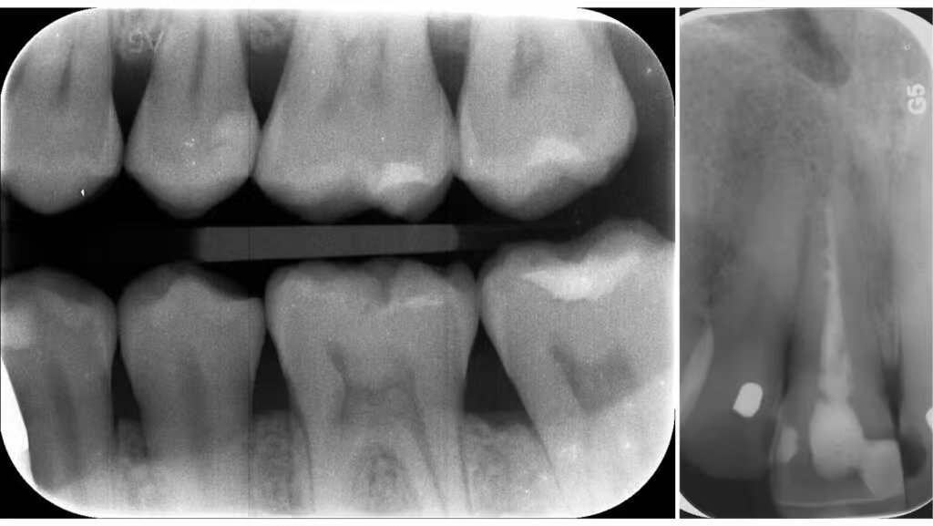
These are 2D X-rays, taken on the inside of the mouth. They capture detailed images of individual teeth, their roots, and the surrounding bone structure. Dentists employ intraoral X-rays to identify tooth decay, bone loss, and dental infections. They are further categorized into bite-wing and periapical X-rays.
- Bite-wing X-rays – These X-rays capture the upper and lower teeth in a single image. This allows the dentist to examine the crowns of the teeth, tooth contacts, and detect decay, gum disease, or bone loss.
- Periapical X-rays – These are small film X-rays taken inside the mouth to capture individual teeth from root to crown. They are useful for detecting tooth decay, bone levels, and root conditions. They provide a comprehensive view of an entire tooth, including surrounding bone, aiding in the diagnosis of abscesses, fractures, and impacted teeth.
Extraoral X-rays
Extraoral X-rays are taken from the outside of the mouth or face, and capture images of the jaw, skull, and facial structures. They provide a broader perspective and are often used to diagnose more complex dental conditions.
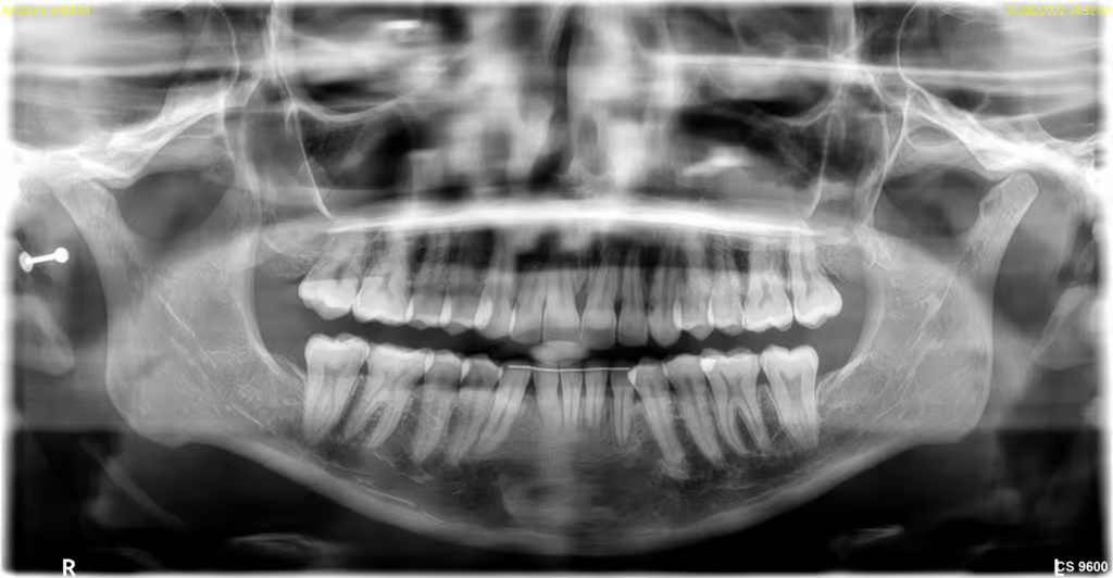
Panoramic X-rays, one of the common extraoral X-rays, is a 2D X-ray and provides a wide view of the entire mouth. This includes the upper and lower jaws, teeth, and temporomandibular joints (TMJ). They are useful for evaluating overall oral health and detecting conditions like impacted teeth, jaw abnormalities, and tumors.
Another type of extraoral X-ray is cone-beam computed tomography (CBCT), which produces three-dimensional images. CBCT X-rays are particularly useful for implant planning, evaluating the jawbone structure, and diagnosing conditions such as tumors or cysts. More about CBCT below.
2D and 3D X-Rays
Both 2D and 3D X-rays play crucial roles in dentistry, but they serve different purposes. 2D X-rays are useful for routine examinations and common dental conditions. 3D X-rays offer significant advantages over 2D X-rays in terms of diagnostic accuracy and treatment planning.
2D X-Rays
2-Dimensional (2D) X-rays, also known as traditional dental radiographs or X-rays, provide a 2D image of the teeth and surrounding structures. 2D X-rays are commonly used for detecting dental caries (cavities), assessing the periodontal condition (gum disease), and evaluating the root structures of teeth. They can also be used for routine dental examinations and screenings.
Types of 2D X-rays include:
- Intraoral Periapical X-rays
- Bitewing X-rays
- Panoramic X-rays
3D X-rays
Cone Beam Computed Tomography (CBCT)
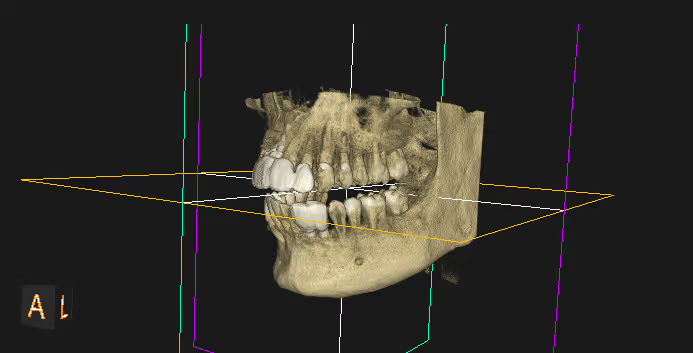
CBCT X-rays (3D X-rays) produce 3D images by rotating a cone-shaped X-ray beam around the patient’s head. The technology captures a series of X-ray images from different angles, which are then reconstructed into a 3D image using specialized software. This provides a three-dimensional view of the teeth, jawbone, and surrounding structures.
With a more comprehensive view of the oral structures, dentists are better able to assess the bone quality, detect abnormalities, evaluate the position of impacted teeth, plan for dental implant placement and orthodontic treatment, assess the airway for sleep apnea, and analyze temporomandibular joint (TMJ) disorders. Additionally, they can view cross-sections of the teeth, jawbone, and surrounding tissues, and enable precise measurements, which can be crucial in complex dental procedures.
3D Dental Scans
3 Dimensional (3D) scans provide a more comprehensive and accurate assessment of the oral structures, enabling precise treatment planning for complex cases.
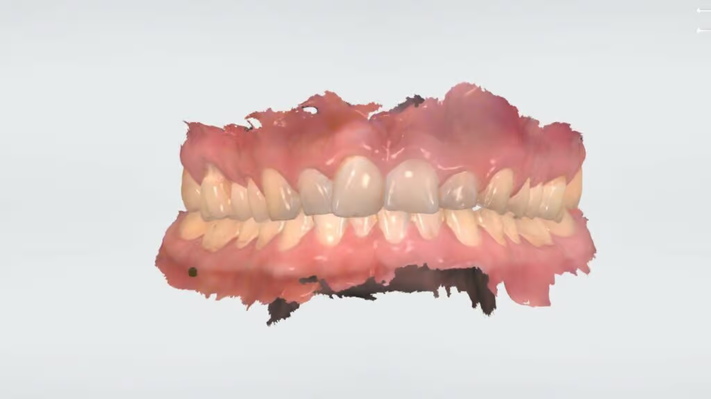
3D Intraoral Scans
A 3D intraoral scan is a dental imaging technology that allows us to create highly accurate digital models of a patient’s teeth and oral structures. By using a handheld wand equipped with a small camera, we capture multiple images of the patient’s mouth from various angles. These images are then combined to create a detailed three-dimensional representation of the teeth and surrounding tissues. This advanced technology eliminates the need for messy traditional dental impressions and provides a more comfortable experience for the patient. The 3D intraoral scan is used for various dental procedures, including orthodontic treatment planning, prosthodontics, and implant dentistry.
Once we have had a consultation, we can determine the specific clinical needs of each patient and weigh the advantages and limitations of each type of scan. We can then recommend the X-rays or scans that would best assist us in formulating your treatment plan.
And if you need any additional information, don’t hesitate to contact us.